Vector Lab代理Vector® TrueVIEW® Autofluorescence Quenching Kit with DAPI

Vector Labs代理,欢迎访问Vector Labs官网或者咨询我们获取相关Vector Labs产品信息以及报价。
Vector Labs 公司是世界著名的生物学检测试剂公司,七十年代其在全球首推的生物素-亲和素系统引领了生物学检测方法的重大革命,该系统被普遍视为目前最灵敏、最可靠与最有效的染色系统,并被广泛应用于免疫组织化学、免疫电镜、原位杂交与凝集素化学中。该系统仍在不断地发展,以满足广大研究人员的各种不同要求。Vector 公司还是世界上凝集素产品的主要供应商,并提供大量的新产品,如酶底物、神经元示踪剂及蛋白质、糖类与核酸的标记、分离与检测试剂。

Vector® TrueVIEW® Autofluorescence Quenching Kit with DAPI

SP-8500-15

SKU Unit Size Price Qty
SP-8500-15 15 ml

To request a quote for products:

  1. Click “Contact Us” in the header bar above;
  2. Click “Customer Service”;
  3. Complete the form and provide the following information in the “Comments” section: note you would like a quote, item number (SKU) and quantity;
  4. Click “Submit”.

Vector® TrueVIEW® Autofluorescence Quenching Kit with DAPI

Description
Specifications
Kit Components
Documents
Product FAQs
Technical Information

Description

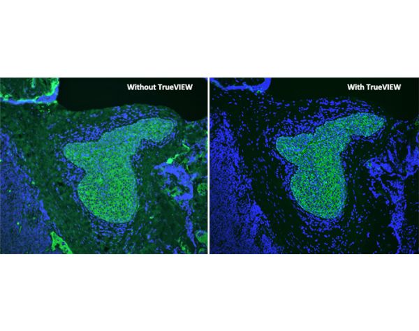
The TrueVIEW Autofluorescence Quenching Kit with DAPI provides a novel way to diminish unwanted autofluorescence from non-lipofuscin sources and dramatically improve signal-to-noise ratio. It removes unwanted autofluorescence in tissue sections due to aldehyde fixation, red blood cells, and structural elements such as collagen and elastin. The quenching action of the kit reagents provides a clear, unambiguous, “true view” localization of the target antigen.

Features:

  • Effective on problematic tissues such as kidney and spleen
  • Easy-to-use, one-step method
  • Compatible with a wide selection of fluorophores
  • Compatible with standard epifluorescence and confocal laser microscopes
  • Mounting medium contains DAPI for counterstaining in one step


One kit is sufficient to treat approximately 100 to 150 tissue sections. 

Specifications

More Information
Unit Size 15 ml
Blocking Action Autofluorescence

Kit Components

Kit Contents:

  • TrueVIEW Reagent A, 5 ml
  • TrueVIEW Reagent B, 5 ml
  • TrueVIEW Reagent C, 5 ml
  • VECTASHIELD® Vibrance™ Antifade Mounting Medium with DAPI, 2 ml

Documents

  • Safety Data Sheet
  • TrueVIEW® Application Note
  • User Guide
  • Download CoA
  • Datasheet

Product FAQs

Why is the TrueVIEW™ Autofluorescence Quenching reagent applied after completion of the IF assay and not at the start of the procedure?

During product development, applying the TrueVIEW™ Quenching reagent at the end of our standard IF procedure yielded the most optimal reduction in autofluorescence signal. The reagent is retained on the tissue at the time of mounting, allowing for extended quenching action, with little to no effect on the specific fluorescent signal. However, in applications with very brief staining procedures, such as a primary antibody directly conjugated with a fluorophore, application of TrueVIEW™ Quenching reagent may be just as effective at the start of the procedure.

Do you have any published references describing the use of TrueVIEW Autofluorescence Quenching Kit?

Yes, there are a number of published references describing the use of TrueVIEW Autofluorescence Quenching Kit in the scientific literature. Please refer to a partial list of these publications in the Technical Information section of the product detail page for SP-8400.

Is TrueVIEW effective against lipofuscin derived autofluorescence?

The working solution of TrueVIEW works in an electrostatic manner to greatly reduce or eliminate fluorescence in tissue sections induced through the use of an aldehyde based fixative. TrueVIEW is also effective at reducing fluorescence from tissue components such as collagen, elastin and red blood cells. TureVIEW does not work to reduce autofluorescence due to lipofuscin.

How long can I store the TrueVIEW working solution?

Once made up, the working solution of TrueVIEW can be stored either on the bench top or in the fridge (2-8C) fro about 48 hours (2 days) withut loss of activity or function. Following this time we would suggest discarding unused working solution and make fresh solution as required.

My tissue sections turned blue when I applied TrueVIEW. Is this supposed to happen?

Yes, the working solution is a blue color that does “stain” the tissue section blue. This indicates an active and appropriate chemical reaction is occurring. The blue stain on the section does not fluoresce and does not interfere with the immunofluorescence application.

What mounting media can I use with TrueVIEW?

Both TrueVIEW products, SP-8400 and SP-8500, are supplied with 2 mL of VECTASHIELD Vibrance antifade mounting media. We have found that the mounting media does play a crucial role in maintaining the high signal to noise ratio when using TrueVIEW. VECTASHIELD Vibrance is included therefore as a critical component of the kit and it is recommended to use the media supplied for optimal results. At this time we do not have sufficient data to confidently recommend the use of other vendors mounting media with TrueVIEW. Substitution of VECTASHIELD Vibrance with another mounting media may result in less than satisfactory results.

For the wash step after applying the TrueVIEW quenching reagent, can I use buffers other than PBS or detergents?

No, we have found that the TrueVIEW reagent lifts off the tissue using TBS or HEPES buffer. Detergents are incompatible.

Technical Information

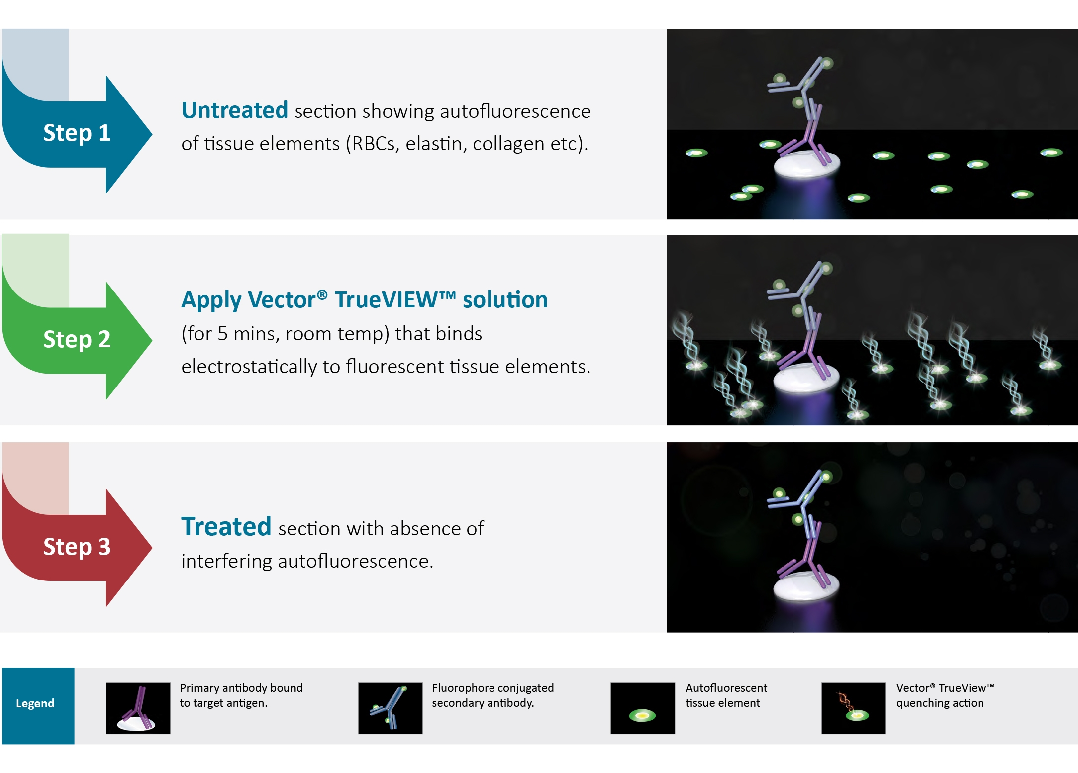
Tissue autofluorescence often occurs with aldehyde fixation or from inherent native tissue components (collagen, elastin, and red blood cells). The extent and intensity of autofluorescence background frequently makes it difficult or impossible to distinguish specific signals in immunofluorescence applications.

Most methods for reduction of tissue autofluorescence act primarily on lipofuscin granules, and are not broadly effective against the most common sources of autofluorescence targeted by TrueVIEW Quencher.

Current methods for reducing autofluorescence primarily include “home brew” concoctions such as sodium borohydride and other ink-based products. These methods are essentially ineffective against aldehyde induced autofluorescence.  In contrast, Vector TrueVIEW reagent binds to hydrophilic compounds and effectively quenches endogenous autofluorescence. 
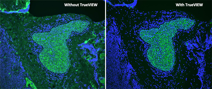
Vector® TrueVIEW® Autofluorescence Quenching Kit with DAPI

Human tonsil (FFPE); Section stained for AE1/AE3 using fluorescein label (green). Treated with TrueVIEW and mounted with VECTASHIELD Vibrance Antifade Mounting Medium with DAPI (nuclei blue). 


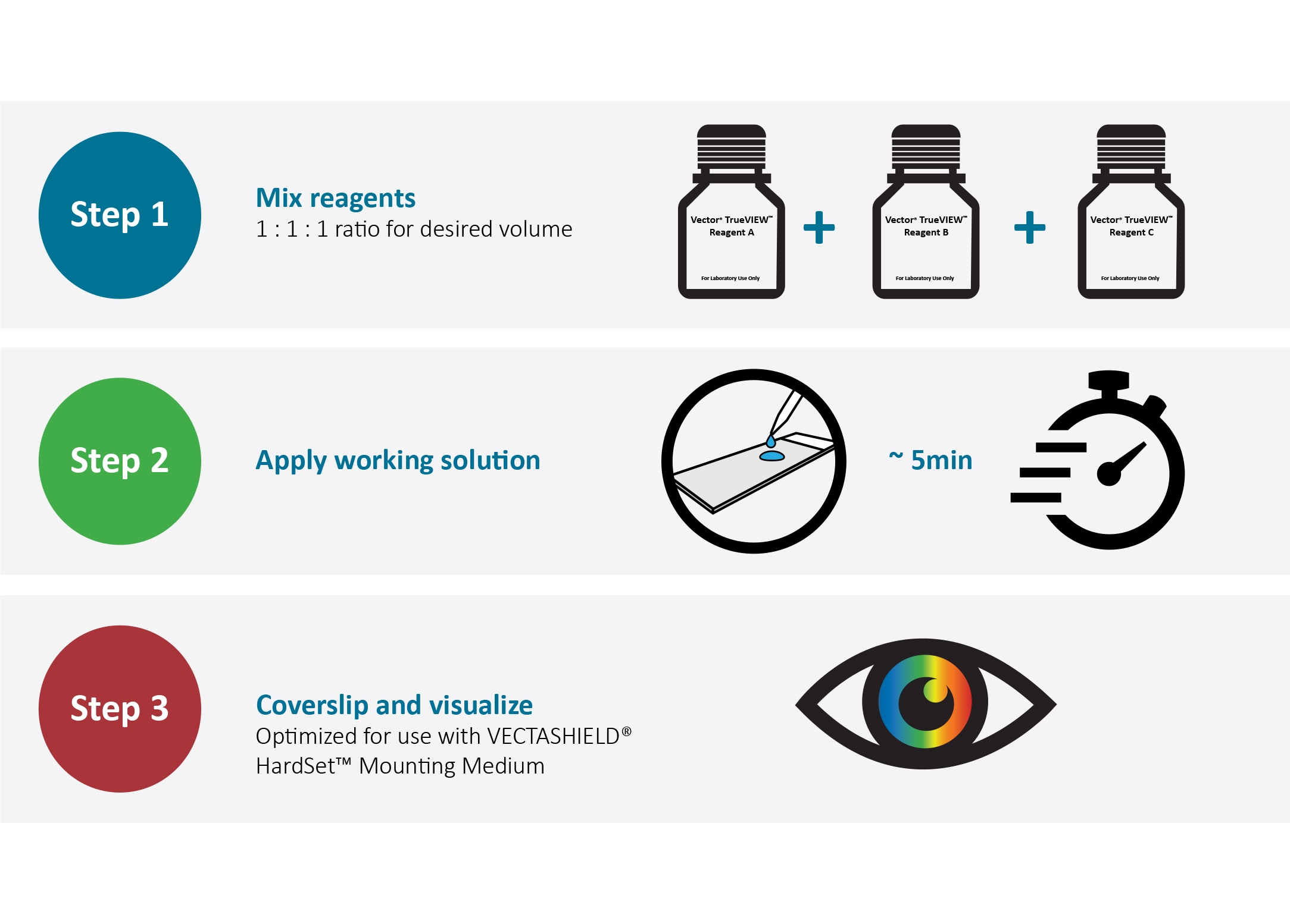
Easy To Apply

Following completion of the IF staining procedure:

Vector® TrueVIEW® Autofluorescence Quenching Kit with DAPI


Mode of Action

Following completion of the IF staining procedure:

Vector® TrueVIEW® Autofluorescence Quenching Kit with DAPI


Signal to Noise Optimization

The primary antibody and detection reagents should be optimized (titered) in conjunction with Vector TrueVIEW Quenching Kit to achieve maximum signal to noise. 

Vector® TrueVIEW® Autofluorescence Quenching Kit with DAPI


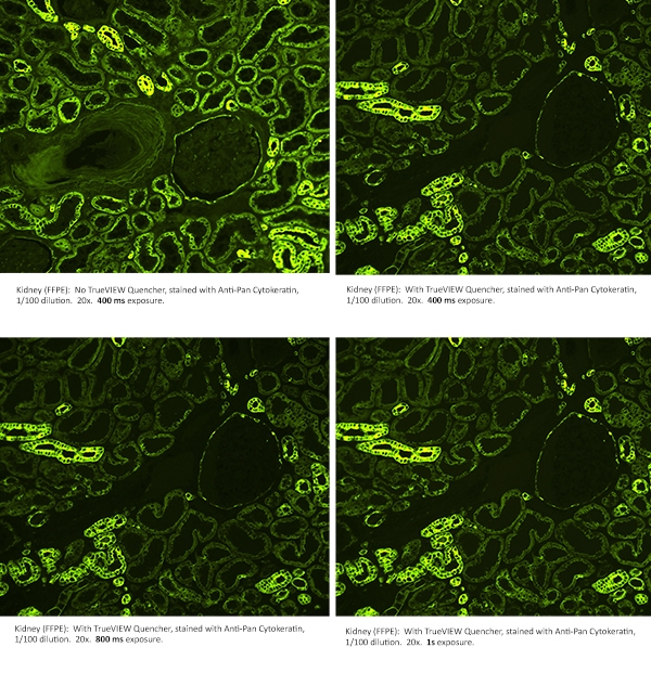
Optimization of Exposure

Increasing exposure times may be necessary to achieve optimal image acquisition for TrueVIEW Quencher treated slides.

Vector® TrueVIEW® Autofluorescence Quenching Kit with DAPI


Comparison Between Vector TrueVIEW Reagent and Other Autofluorescence Reducing Agents

We compared the effectiveness of Vector TrueVIEW quenching action in parallel with other commercially available autofluorescence reducing products and “home brew” reagents, on serial sections of formalin-fixed, paraffin-embedded human pancreas visualized using a standard fluorescein (green) filter. No specific immunofluorescence staining was conducted. The images below highlight our results. All images were acquired under identical conditions (including microscope objective and exposure times). 

Vector® TrueVIEW® Autofluorescence Quenching Kit with DAPI


Customer Testimonials 

Vector® TrueVIEW® Autofluorescence Quenching Kit with DAPI

Home All Products Blocking Reagents What’s New Autofluorescence Quenching Kits Vector® TrueVIEW® Autofluorescence Quenching Kit with DAPI

Vector Lab代理VECTASHIELD® Antifade Mounting Medium with DAPI

Vector Labs代理,欢迎访问Vector Labs官网或者咨询我们获取相关Vector Labs产品信息以及报价。
Vector Labs 公司是世界著名的生物学检测试剂公司,七十年代其在全球首推的生物素-亲和素系统引领了生物学检测方法的重大革命,该系统被普遍视为目前最灵敏、最可靠与最有效的染色系统,并被广泛应用于免疫组织化学、免疫电镜、原位杂交与凝集素化学中。该系统仍在不断地发展,以满足广大研究人员的各种不同要求。Vector 公司还是世界上凝集素产品的主要供应商,并提供大量的新产品,如酶底物、神经元示踪剂及蛋白质、糖类与核酸的标记、分离与检测试剂。

VECTASHIELD® Antifade Mounting Medium with DAPI

H-1200-10

Still using the original VECTASHIELD with DAPI?

Try our upgraded product VECTASHIELD® PLUS Antifade Mounting Medium with DAPI

SKU Unit Size Price Qty
H-1200-10 10 ml

To request a quote for products:

  1. Click “Contact Us” in the header bar above;
  2. Click “Customer Service”;
  3. Complete the form and provide the following information in the “Comments” section: note you would like a quote, item number (SKU) and quantity;
  4. Click “Submit”.

VECTASHIELD® Antifade Mounting Medium with DAPI

Description
Specifications
Documents
Product FAQs
Related Products
Citations
Technical Information

Description

VECTASHIELD Antifade Mounting Medium with DAPI is a unique, stable formula for preserving fluorescence. This medium inhibits rapid photobleaching of fluorescent proteins and fluorescent dyes. VECTASHIELD Mounting Medium with DAPI does not solidify, but remains a liquid on the slide and can be stored without sealing.

Features:

  • Ideal refractive index
  • Ready to use
  • No warming necessary
  • Can be stored without sealing for long term analysis
  • Easy to use

Specifications

More Information
Unit Size 10 ml
Applications Immunofluorescence, In situ hybridization, Cellular Imaging
Mounting Aqueous (Non-Hardening)
Antifade Yes
Counterstain DAPI (blue fluorescence)
Cellular Stain Nucleus

Documents

  • VECTASHIELD Fluorochrome Compatibility
  • User Guide
  • In Situ Hybridization Detection Protocol
  • Safety Data Sheet
  • Download CoA
  • Datasheet

Product FAQs

Can VECTASHIELD® be used for super resolution microscopy?

Of the many variations of super resolution microscopy (SRM) currently being used, investigators have found the properties of product H-1000 (VECTASHIELD), to be advantageous in stochastic optical reconstruction microscopy (STORM) and structured illumination microscopy (SIM). Refer to the following published references for further information: 1) Olivier, N., et al (2013) Simple buffers for 3D STORM microscopy. Biochemical Optics Express 4:885-899. 2) Wegel., et al (2016) Imaging cellular structuires in super-resolution with SIM, STED and localisation microscopy: A practical comparison. Scientific Reports 6:27290.

Does VECTASHIELD contain glycerol?

All of the VECTASHIELD formats we currently offer which includes regular non-setting VECTASHIELD, PLUS, HardSet and Vibrance formulations each contain a percentage of glycerol.

Do I need to dehydrate the tissue sections before applying VECTASHIELD?

No form of tissue dehydration (e.g. air drying or ethanol exposure) is required nor recommended when applying VECTASHIELD. From our experience, the most optimal antifade actions of VECTASHIELD are obtained when the preparation is removed from the final buffer/water rinse, kept slightly wet/moist and then coverslipped with a small volume (25-50 uL) of VECTASHIELD.

Do I need to seal the perimeter of the coverslip after I apply VECTASHIELD?

That depends upon how long you wish to retain the slides and which VECTASHIELD formulation you are using. If you are using one of our non-setting VECTASHIELD products such as H-1000/H-1200 or H-1900/H-2000, then we suggest sealing the coverslip with plastic sealant or nail polish if you intend to keep the slides beyond a day or so. If you are using one of our setting/curing formulations such as VECTASHIELD HardSet or Vibrance, then in most cases when using thin cut (<10 um) tissue sections or cell monolayers, no sealing of the coverslip is requuired.

Related Products

VECTASHIELD® Antifade Mounting Medium with DAPI

VECTASHIELD® PLUS Antifade Mounting Medium Catalog #: H-1900

VECTASHIELD® Antifade Mounting Medium with DAPI

VECTASHIELD® PLUS Antifade Mounting Medium with DAPI Catalog #: H-2000

VECTASHIELD® Antifade Mounting Medium with DAPI

VECTASHIELD® Vibrance™ Antifade Mounting Medium Catalog #: H-1700

VECTASHIELD® Antifade Mounting Medium with DAPI

VECTASHIELD® Vibrance™ Antifade Mounting Medium with DAPI Catalog #: H-1800

VECTASHIELD® Antifade Mounting Medium with DAPI

VECTASHIELD® Antifade Mounting Medium Catalog #: H-1000-10

VECTASHIELD® Antifade Mounting Medium with DAPI

VECTASHIELD® Antifade Mounting Medium with Propidium Iodide (PI) Catalog #: H-1300-10

VECTASHIELD® Antifade Mounting Medium with DAPI

VECTASHIELD® HardSet™ Antifade Mounting Medium Catalog #: H-1400-10

VECTASHIELD® Antifade Mounting Medium with DAPI

VECTASHIELD® HardSet™ Antifade Mounting Medium with DAPI Catalog #: H-1500-10

VECTASHIELD® Antifade Mounting Medium with DAPI

VECTASHIELD® HardSet™ Antifade Mounting Medium with Phalloidin Catalog #: H-1600-10

Citations

VECTASHIELD® Antifade Mounting Medium with DAPI Powered by Bioz See more details on Bioz

Technical Information

VECTASHIELD Mounting Media are compatible with a wide array of fluorochromes, enzymatic substrates, and fluorescent proteins. For more information about the compatibility of VECTASHIELD products, please consult the VECTASHIELD® Mounting Medium Compatibility table.

All formulations of VECTASHIELD Antifade Mounting Media are available with or without the counterstain DAPI (4', 6-diamidino-2-phenylindole), which produces a blue fluorescence when bound to DNA with excitation at about 360 nm and emission at 460 nm.  If desired, the concentration of DAPI can be reduced by mixing with the corresponding VECTASHIELD Mounting Medium (without DAPI).

If desired, coverslips can be sealed around the perimeter with nail polish or a plastic sealant. Mounted slides should be stored at 4 °C, protected from light.

The refractive index for VECTASHIELD Mounting Medium is 1.45.

Publications

R.J. Florijn, et al. 1995. Analysis of antifading reagents for fluorescence microscopy. Cytometry. doi: 10.1002/cyto.990190213

VECTASHIELD Mounting Medium Antifade Comparison

Other manufacturers measure the antifade properties of their mountants using labeled microspheres or arrayed spots. Vector Labs prefers to measure antifade properties of VECTASHIELD mountants using frozen tissue sections immunohistochemically stained with fluorescently labeled secondary antibodies. Antifade capability is then measured using a 40x objective with real time imaging over 30 seconds of continuous exposure to the excitation illumination. Individual intensity measurements are recorded from 6 separate labeled regions and the average is calculated. The intensity after 30 second exposure is expressed as a percentage of the intensity at zero time. The values for PG are taken from the manufacturer s published results.

Product Counterstain Catalog Number Unit Size Hardening Refractive Index

VECTASHIELD Antifade Mounting Medium

None

H-1000

10 ml

No

1.45

DAPI

H-1200

10 ml

No

1.45

PI

H-1300

10 ml

No

1.45

VECTASHIELD PLUS Antifade Mounting Medium

None

H-1900

2 ml, 10 ml

No

1.45

DAPI

H-2000

2 ml, 10 ml

No

1.45

VECTASHIELD HardSet Antifade Mounting Medium

None

H-1400

10 ml

Yes

1.36 (initial)
1.46 (cured)

DAPI

H-1500

10 ml

Yes

1.36 (initial)
1.46 (cured)

TRITC-Phalloidin

H-1600

10 ml

Yes

1.36 (initial)
1.46 (cured)

VECTASHIELD Vibrance Antifade Mounting Medium

None

H-1700

2 ml, 10 ml

Yes

1.38 (initial)
1.47 (cured)

DAPI

H-1800

2 ml, 10 ml

Yes

1.38 (initial)
1.47 (cured)

Additional Properties of VECTASHIELD Antifade Mounting Medium

VECTASHIELD® Antifade Mounting Medium with DAPI

Home All Products VECTASHIELD Antifade Mounting Media (IF) VECTASHIELD® Antifade Mounting Medium with DAPI

Vector Lab代理Horse Anti-Mouse/Rabbit/Goat IgG Antibody (H+L) (Universal Pan-Specific ), Biotinylated

Vector Labs代理,欢迎访问Vector Labs官网或者咨询我们获取相关Vector Labs产品信息以及报价。
Vector Labs 公司是世界著名的生物学检测试剂公司,七十年代其在全球首推的生物素-亲和素系统引领了生物学检测方法的重大革命,该系统被普遍视为目前最灵敏、最可靠与最有效的染色系统,并被广泛应用于免疫组织化学、免疫电镜、原位杂交与凝集素化学中。该系统仍在不断地发展,以满足广大研究人员的各种不同要求。Vector 公司还是世界上凝集素产品的主要供应商,并提供大量的新产品,如酶底物、神经元示踪剂及蛋白质、糖类与核酸的标记、分离与检测试剂。

Horse Anti-Mouse/Rabbit/Goat IgG Antibody (H+L) (Universal Pan-Specific ), Biotinylated

BA-1300-2.2

SKU Unit Size Price Qty
BA-1300-2.2 2.2 ml

To request a quote for products:

  1. Click “Contact Us” in the header bar above;
  2. Click “Customer Service”;
  3. Complete the form and provide the following information in the “Comments” section: note you would like a quote, item number (SKU) and quantity;
  4. Click “Submit”.

Horse Anti-Mouse/Rabbit/Goat IgG Antibody (H+L) (Universal Pan-Specific ), Biotinylated

Description
Specifications
Documents
Related Products
Citations
Technical Information

Description

This pan-specific, biotinylated universal secondary antibody is specially formulated to recognize IgG (H+L) primary antibodies from mouse, rabbit, or goat. This mixture of biotinylated secondary antibodies is produced in horse, and provides the same high sensitivity and low background as that obtained with our species-specific biotinylated anti-mouse IgG (H+L), biotinylated anti-rabbit IgG (H+L), or biotinylated anti-goat IgG (H+L). Use of this pan-specific secondary antibody provides added convenience and is adaptable to automated instrument staining.

Features:

  • Recognizes both heavy and light chains (H+L) 
  • Biotinylated to ensure the maximum degree of labeling without compromising the specificity or affinity of the antibody 
  • Can be used for tissue and cell staining, ELISAs, and blots 
  • Included in the VECTASTAIN® Universal Quick kits
  • Supplied as 2.2 ml of solution
  • Recommended dilution for most applications is 1:20
  • This pan-specific, biotinylated “universal” secondary antibody is specially formulated to recognize IgG (H+L) primary antibodies from mouse, rabbit, or goat.

Note: This antibody has significant cross-reactivity for rodent, rabbit, goat, sheep and bovine IgG and is not recommended for use in staining tissues of these species.

Note: This product is the same biotinylated secondary antibody included in the VECTASTAIN® Universal Quick Kit, PK-8800, but is not the same as that provided in the VECTASTAIN® Elite ABC Kit (Universal), PK-6200.

Specifications

More Information
Unit Size 2.2 ml
Applications Immunohistochemistry / Immunocytochemistry, Immunofluorescence, In situ hybridization, Blotting Applications, Elispot, ELISAs
Concentration 0.91 mg/ml
Recommended Storage 2-8 °C; Store frozen for long term storage
Solution 10 mM sodium phosphate, pH 7.8, 0.15 M NaCl, 0.08% sodium azide, 3 mg/ml bovine serum albumin.
Recommended Usage NOTE:This product is a specially prepared biotinylated secondary antibody which recognizes primary antibodies made in mouse, rabbit or goat.This antibody should not be used on tissues from mouse, rabbit or goat or from animals related immunologically to these species.Significant background due to crossreactivity with endogenous immunoglobulins may result from its inappropriate use.This product is the same as that included in the VECTASTAIN® Universal Quick Kit (Cat. No. PK-8800)but is not the same as the antibody included in the VECTASTAIN® Universal Elite® ABC Kit. If this biotinylated antibody is to be used in a 30 minute incubation, dispense 4 drops into 5 ml of phosphate buffered saline (PBS) containing 1.5% normal horse serum. If this biotinylated antibody is to be used in a 10 minute incubation, dispense 1 drop per ml of PBS containing 5-10% normal horse serum.
Target Species Universal (Mouse/Rabbit/Goat)
Conjugate Biotinylated
Host Species Horse
Format Concentrate

Documents

  • Safety Data Sheet
  • Download CoA
  • Datasheet

Related Products

Horse Anti-Mouse/Rabbit/Goat IgG Antibody (H+L) (Universal Pan-Specific ), Biotinylated

Fluorescein Avidin DCS, Cell Sorting Grade Catalog #: A-2011-1

Horse Anti-Mouse/Rabbit/Goat IgG Antibody (H+L) (Universal Pan-Specific ), Biotinylated

Texas Red® Avidin DCS, Cell Sorting Grade Catalog #: A-2016-1

Horse Anti-Mouse/Rabbit/Goat IgG Antibody (H+L) (Universal Pan-Specific ), Biotinylated

VECTASTAIN® ABC-AP Kit, Alkaline Phosphatase (Standard) Catalog #: AK-5000

Horse Anti-Mouse/Rabbit/Goat IgG Antibody (H+L) (Universal Pan-Specific ), Biotinylated

VECTASTAIN® ABC-HRP Kit, Peroxidase (Standard) Catalog #: PK-4000

Horse Anti-Mouse/Rabbit/Goat IgG Antibody (H+L) (Universal Pan-Specific ), Biotinylated

VECTASTAIN® Elite ABC-HRP Kit, Peroxidase (Standard) Catalog #: PK-6100

Horse Anti-Mouse/Rabbit/Goat IgG Antibody (H+L) (Universal Pan-Specific ), Biotinylated

Streptavidin, DyLight® 488 Catalog #: SA-5488-1

Horse Anti-Mouse/Rabbit/Goat IgG Antibody (H+L) (Universal Pan-Specific ), Biotinylated

Streptavidin, DyLight® 549 Catalog #: SA-5549-1

Horse Anti-Mouse/Rabbit/Goat IgG Antibody (H+L) (Universal Pan-Specific ), Biotinylated

Streptavidin, DyLight® 594 Catalog #: SA-5594-1

Horse Anti-Mouse/Rabbit/Goat IgG Antibody (H+L) (Universal Pan-Specific ), Biotinylated

Streptavidin, DyLight® 649 Catalog #: SA-5649-1

Horse Anti-Mouse/Rabbit/Goat IgG Antibody (H+L) (Universal Pan-Specific ), Biotinylated

Streptavidin, Peroxidase, R.T.U. Catalog #: SA-5704-100

Citations

Horse Anti-Mouse/Rabbit/Goat IgG Antibody (H+L) (Universal Pan-Specific ), Biotinylated Powered by Bioz See more details on Bioz

Technical Information

Vector Laboratories affinity-purified antibodies are of unmatched quality. These antibodies are prepared using proprietary immunization schedules that produce high affinity antibodies. The antibodies are then purified by affinity chromatography, and cross-reactivities that are likely to interfere with specific labeling are removed by solid phase adsorption techniques. The biotinylated secondary antibodies are conjugated to ensure the maximum degree of labeling without compromising the specificity or affinity of the antibody.


Home All Products Biotinylated Secondary Antibodies Horse Anti-Mouse/Rabbit/Goat IgG Antibody (H+L) (Universal Pan-Specific ), Biotinylated

Vector Lab代理Goat Anti-Human IgE Antibody, epsilon chain specific, Biotinylated

Vector Labs代理,欢迎访问Vector Labs官网或者咨询我们获取相关Vector Labs产品信息以及报价。
Vector Labs 公司是世界著名的生物学检测试剂公司,七十年代其在全球首推的生物素-亲和素系统引领了生物学检测方法的重大革命,该系统被普遍视为目前最灵敏、最可靠与最有效的染色系统,并被广泛应用于免疫组织化学、免疫电镜、原位杂交与凝集素化学中。该系统仍在不断地发展,以满足广大研究人员的各种不同要求。Vector 公司还是世界上凝集素产品的主要供应商,并提供大量的新产品,如酶底物、神经元示踪剂及蛋白质、糖类与核酸的标记、分离与检测试剂。

Goat Anti-Human IgE Antibody, epsilon chain specific, Biotinylated

BA-3040-.5

SKU Unit Size Price Qty
BA-3040-.5 0.5 mg

To request a quote for products:

  1. Click “Contact Us” in the header bar above;
  2. Click “Customer Service”;
  3. Complete the form and provide the following information in the “Comments” section: note you would like a quote, item number (SKU) and quantity;
  4. Click “Submit”.

Goat Anti-Human IgE Antibody, epsilon chain specific, Biotinylated

Description
Specifications
Documents
Related Products
Citations
Technical Information

Description

Biotinylated Goat Anti-Human IgE, epsilon chain specific, secondary antibody is used in an avidin-biotin or streptavidin-biotin detection system. This epsilon chain specific antibody has virtually no cross-reactivity with other immunoglobulin classes or other heavy or light chains. 

Features:

  • Biotinylated to ensure the maximum degree of labeling without compromising the specificity or affinity of the antibody 
  • Can be used for flow cytometry or used for tissue staining 

Specifications

More Information
Unit Size 0.5 mg
Applications Immunohistochemistry / Immunocytochemistry, Immunofluorescence, In situ hybridization, Blotting Applications, Elispot, ELISAs
Recommended Storage 2-8 °C; Store frozen for long term storage
Recommended Usage Reconstitute by adding 1 ml water.The resulting solution will have the following composition:10 mM sodium phosphate, pH 7.8, 0.15 M NaCl, 0.08% sodium azide, 1 mg/ml bovine serum albumin. The recommended concentration range for use is 2-10 µg/ml.If this biotinylated antibody is to be used in tissues, which may contain cross-reacting endogenous immunoglobulins, dilution of this biotinylated antibody may be made in buffers containing 2% normal serum from the same species as the tissue.
Target Species Human
Conjugate Biotinylated
Host Species Goat
Format Lyophilized

Documents

  • Safety Data Sheet
  • Download CoA
  • Datasheet

Related Products

Goat Anti-Human IgE Antibody, epsilon chain specific, Biotinylated

Fluorescein Avidin DCS, Cell Sorting Grade Catalog #: A-2011-1

Goat Anti-Human IgE Antibody, epsilon chain specific, Biotinylated

Texas Red® Avidin DCS, Cell Sorting Grade Catalog #: A-2016-1

Goat Anti-Human IgE Antibody, epsilon chain specific, Biotinylated

VECTASTAIN® ABC-AP Kit, Alkaline Phosphatase (Standard) Catalog #: AK-5000

Goat Anti-Human IgE Antibody, epsilon chain specific, Biotinylated

VECTASTAIN® ABC-HRP Kit, Peroxidase (Standard) Catalog #: PK-4000

Goat Anti-Human IgE Antibody, epsilon chain specific, Biotinylated

VECTASTAIN® Elite ABC-HRP Kit, Peroxidase (Standard) Catalog #: PK-6100

Goat Anti-Human IgE Antibody, epsilon chain specific, Biotinylated

Streptavidin, DyLight® 488 Catalog #: SA-5488-1

Goat Anti-Human IgE Antibody, epsilon chain specific, Biotinylated

Streptavidin, DyLight® 549 Catalog #: SA-5549-1

Goat Anti-Human IgE Antibody, epsilon chain specific, Biotinylated

Streptavidin, DyLight® 594 Catalog #: SA-5594-1

Goat Anti-Human IgE Antibody, epsilon chain specific, Biotinylated

Streptavidin, DyLight® 649 Catalog #: SA-5649-1

Goat Anti-Human IgE Antibody, epsilon chain specific, Biotinylated

Streptavidin, Peroxidase, R.T.U. Catalog #: SA-5704-100

Citations

Goat Anti-Human IgE Antibody, epsilon chain specific, Biotinylated Powered by Bioz See more details on Bioz

Technical Information

Vector Laboratories affinity-purified antibodies are prepared using proprietary immunization schedules that produce high affinity antibodies. The antibodies are then purified by affinity chromatography, and cross-reactivities that are likely to interfere with specific labeling are removed by solid phase adsorption techniques. The biotinylated secondary antibodies are conjugated to ensure the maximum degree of labeling without compromising the specificity or affinity of the antibody. These antibodies are subjected to rigorous quality control assays and can be used for tissue and cell staining, ELISAs, and blots.

Biotinylated Goat Anti-Human IgE, epsilon chain specific, is supplied in lyophilized form and can be reconstituted with 1 ml water. With some exceptions the recommended dilution for most applications is 1:100.

This antibody is affinity-purified and treated by negative solid-phase adsorption to remove cross-reacting antibodies. This chain specific antibody has virtually no cross-reactivity with other immunoglobulin classes or other heavy or light chains. This biotinylated conjugate can be employed in flow cytometry or used for tissue staining.

Home All Products Biotinylated Secondary Antibodies Goat Anti-Human IgE Antibody, epsilon chain specific, Biotinylated

Vector Lab代理Goat Anti-Chicken IgY Antibody (H+L), Biotinylated

Vector Labs代理,欢迎访问Vector Labs官网或者咨询我们获取相关Vector Labs产品信息以及报价。
Vector Labs 公司是世界著名的生物学检测试剂公司,七十年代其在全球首推的生物素-亲和素系统引领了生物学检测方法的重大革命,该系统被普遍视为目前最灵敏、最可靠与最有效的染色系统,并被广泛应用于免疫组织化学、免疫电镜、原位杂交与凝集素化学中。该系统仍在不断地发展,以满足广大研究人员的各种不同要求。Vector 公司还是世界上凝集素产品的主要供应商,并提供大量的新产品,如酶底物、神经元示踪剂及蛋白质、糖类与核酸的标记、分离与检测试剂。

Goat Anti-Chicken IgY Antibody (H+L), Biotinylated

BA-9010-1.5

SKU Unit Size Price Qty
BA-9010-1.5 1.5 mg

To request a quote for products:

  1. Click “Contact Us” in the header bar above;
  2. Click “Customer Service”;
  3. Complete the form and provide the following information in the “Comments” section: note you would like a quote, item number (SKU) and quantity;
  4. Click “Submit”.

Goat Anti-Chicken IgY Antibody (H+L), Biotinylated

Description
Specifications
Documents
Related Products
Citations
Technical Information

Description

The biotinylated goat anti-chicken IgY secondary antibody is used in an avidin-biotin or streptavidin-biotin detection system. These antibodies can be used for tissue and cell staining, ELISAs, and blots.


Features:

  • Suitable for use with chicken IgY and IgG primary antibodies
  • Recognizes both heavy and light chains (H+L) 
  • Biotinylated to ensure the maximum degree of labeling without compromising the specificity or affinity of the antibody 

Specifications

More Information
Unit Size 1.5 mg
Applications Immunohistochemistry / Immunocytochemistry, Immunofluorescence, In situ hybridization, Blotting Applications, Elispot, ELISAs
Concentration 1.5 mg active conjugate/ml
Recommended Storage 2-8 °C; Store frozen for long term storage
Solution 10 mM sodium phosphate, pH 7.8, 0.15 M NaCl, 0.08% sodium azide, 3 mg/ml bovine serum albumin.
Recommended Usage The recommended concentration range for use is 2-10 µg/ml.If this biotinylated antibody is to be used in tissues, which may contain cross-reacting endogenous immunoglobulins, dilution of this biotinylated antibody may be made in buffers containing 2% normal serum from the same species as the tissue.
Target Species Chicken
Conjugate Biotinylated
Host Species Goat
Format Concentrate

Documents

  • Safety Data Sheet
  • Download CoA
  • Datasheet

Related Products

Goat Anti-Chicken IgY Antibody (H+L), Biotinylated

Fluorescein Avidin DCS, Cell Sorting Grade Catalog #: A-2011-1

Goat Anti-Chicken IgY Antibody (H+L), Biotinylated

Texas Red® Avidin DCS, Cell Sorting Grade Catalog #: A-2016-1

Goat Anti-Chicken IgY Antibody (H+L), Biotinylated

VECTASTAIN® ABC-AP Kit, Alkaline Phosphatase (Standard) Catalog #: AK-5000

Goat Anti-Chicken IgY Antibody (H+L), Biotinylated

VECTASTAIN® ABC-HRP Kit, Peroxidase (Standard) Catalog #: PK-4000

Goat Anti-Chicken IgY Antibody (H+L), Biotinylated

VECTASTAIN® Elite ABC-HRP Kit, Peroxidase (Standard) Catalog #: PK-6100

Goat Anti-Chicken IgY Antibody (H+L), Biotinylated

Streptavidin, DyLight® 488 Catalog #: SA-5488-1

Goat Anti-Chicken IgY Antibody (H+L), Biotinylated

Streptavidin, DyLight® 549 Catalog #: SA-5549-1

Goat Anti-Chicken IgY Antibody (H+L), Biotinylated

Streptavidin, DyLight® 594 Catalog #: SA-5594-1

Goat Anti-Chicken IgY Antibody (H+L), Biotinylated

Streptavidin, DyLight® 649 Catalog #: SA-5649-1

Goat Anti-Chicken IgY Antibody (H+L), Biotinylated

Streptavidin, Peroxidase, R.T.U. Catalog #: SA-5704-100

Citations

Goat Anti-Chicken IgY Antibody (H+L), Biotinylated Powered by Bioz See more details on Bioz

Technical Information

Vector Laboratories affinity-purified antibodies are of unmatched quality. These antibodies are prepared using proprietary immunization schedules that produce high affinity antibodies. The antibodies are then purified by affinity chromatography, and cross-reactivities that are likely to interfere with specific labeling are removed by solid phase adsorption techniques. The biotinylated secondary antibodies are conjugated to ensure the maximum degree of labeling without compromising the specificity or affinity of the antibody. These antibodies are subjected to rigorous quality control assays and can be used with any avidin or streptavidin detection system for tissue and cell staining, ELISAs, and blots.

Biotinylated Goat Anti-Chicken IgY (H+L) is supplied in solution form. With some exceptions the recommended dilution for most applications is 1:200.

Home All Products Biotinylated Secondary Antibodies Goat Anti-Chicken IgY Antibody (H+L), Biotinylated

Vector Lab代理Goat Anti-Rabbit IgG Antibody (H+L), AMCA

Vector Labs代理,欢迎访问Vector Labs官网或者咨询我们获取相关Vector Labs产品信息以及报价。
Vector Labs 公司是世界著名的生物学检测试剂公司,七十年代其在全球首推的生物素-亲和素系统引领了生物学检测方法的重大革命,该系统被普遍视为目前最灵敏、最可靠与最有效的染色系统,并被广泛应用于免疫组织化学、免疫电镜、原位杂交与凝集素化学中。该系统仍在不断地发展,以满足广大研究人员的各种不同要求。Vector 公司还是世界上凝集素产品的主要供应商,并提供大量的新产品,如酶底物、神经元示踪剂及蛋白质、糖类与核酸的标记、分离与检测试剂。

Goat Anti-Rabbit IgG Antibody (H+L), AMCA

CI-1000-1.5

SKU Unit Size Price Qty
CI-1000-1.5 1.5 mg

To request a quote for products:

  1. Click “Contact Us” in the header bar above;
  2. Click “Customer Service”;
  3. Complete the form and provide the following information in the “Comments” section: note you would like a quote, item number (SKU) and quantity;
  4. Click “Submit”.

Goat Anti-Rabbit IgG Antibody (H+L), AMCA

Description
Specifications
Documents
Related Products
Citations
Technical Information

Description

AMCA Goat Anti-Rabbit IgG Antibody can be used for immunofluorescence and other applications. Optimal F/P ratios have been established for each conjugate to ensure maximum fluorescence with minimal background staining.

Features:

  • Recognizes both heavy and light chains (H+L)
  • Optimally labeled with AMCA to provide the brightest label for fluorescence microscopy
  • Supplied in solution
  • Excitation: 350 nm
  • Emission: 450 nm
  • Color: Blue

Specifications

More Information
Unit Size 1.5 mg
Applications Immunofluorescence, In situ hybridization, Flow Cytometry/Cell Separation
Concentration 1.5 mg active conjugate/ml
Recommended Storage 2-8 °C
Solution 10 mM HEPES, 0.15 M NaCl, pH 7.5, 0.08% sodium azide.
Maximum Emission 448-454 nm.
Maximum Excitation 345-355 nm
Recommended Usage Maximum Emission @ 510-520 nm
Target Species Rabbit
Conjugate AMCA
Color of Fluorescence Blue
Host Species Goat
Format Concentrate

Documents

  • Safety Data Sheet
  • Download CoA
  • Datasheet

Related Products

Goat Anti-Rabbit IgG Antibody (H+L), AMCA

Horse Anti-Rabbit IgG Antibody (H+L), DyLight® 488 Catalog #: DI-1088-1.5

Goat Anti-Rabbit IgG Antibody (H+L), AMCA

Horse Anti-Rabbit IgG Antibody (H+L), DyLight® 594 Catalog #: DI-1094-1.5

Goat Anti-Rabbit IgG Antibody (H+L), AMCA

Goat Anti-Rabbit IgG Antibody (H+L), DyLight® 488 Catalog #: DI-1488-1.5

Goat Anti-Rabbit IgG Antibody (H+L), AMCA

Goat Anti-Rabbit IgG Antibody (H+L), DyLight® 549 Catalog #: DI-1549-1.5

Goat Anti-Rabbit IgG Antibody (H+L), AMCA

Goat Anti-Rabbit IgG Antibody (H+L), DyLight® 594 Catalog #: DI-1594-1.5

Goat Anti-Rabbit IgG Antibody (H+L), AMCA

Goat Anti-Rabbit IgG Antibody (H+L), DyLight® 649 Catalog #: DI-1649-1.5

Goat Anti-Rabbit IgG Antibody (H+L), AMCA

VectaFluor™ Horse Anti-Rabbit IgG, DyLight® 488 Antibody Kit, R.T.U. Catalog #: DI-1788

Goat Anti-Rabbit IgG Antibody (H+L), AMCA

VectaFluor™ Horse Anti-Rabbit IgG, DyLight® 594 Antibody Kit, R.T.U. Catalog #: DI-1794

Goat Anti-Rabbit IgG Antibody (H+L), AMCA

VectaFluor™ Excel Amplified Anti-Rabbit IgG, DyLight® 488 Antibody Kit Catalog #: DK-1488

Goat Anti-Rabbit IgG Antibody (H+L), AMCA

VectaFluor™ Excel Amplified Anti-Rabbit IgG, DyLight® 594 Antibody Kit Catalog #: DK-1594

Goat Anti-Rabbit IgG Antibody (H+L), AMCA

Goat Anti-Rabbit IgG Antibody (H+L), Fluorescein Catalog #: FI-1000-1.5

Goat Anti-Rabbit IgG Antibody (H+L), AMCA

VECTASHIELD® Antifade Mounting Medium Catalog #: H-1000-10

Goat Anti-Rabbit IgG Antibody (H+L), AMCA

VECTASHIELD® HardSet™ Antifade Mounting Medium Catalog #: H-1400-10

Goat Anti-Rabbit IgG Antibody (H+L), AMCA

Goat Anti-Rabbit IgG Antibody (H+L), Texas Red® Catalog #: TI-1000-1.5

Goat Anti-Rabbit IgG Antibody (H+L), AMCA

Goat Anti-Rabbit IgG Antibody (H+L), Biotinylated Catalog #: BA-1000-1.5

Citations

Goat Anti-Rabbit IgG Antibody (H+L), AMCA Powered by Bioz See more details on Bioz

Technical Information

The goat anti-rabbit Ig antibodies are prepared by hyperimmunizing animals in a manner that produces high affinity antibodies. These are then purified by an affinity chromatography procedure designed to remove any low affinity antibodies which may be present. Cross-reactivities that are likely to interfere with specific labeling are removed by solid-phase adsorption techniques. The final product is then subjected to rigorous quality control assays including immunodiffusion, solid-phase enzyme immunoassays, gel electrophoresis and solid-phase binding assays. In preparing the labeled antibodies, great care is taken to ensure the maximum degree of labeling with no alteration in the specificity and affinity of the antibody. The labeled antibody then undergoes a further series of quality control assays, including immunohistochemical analysis.

Home All Products Fluorophore-Conjugated Secondary Antibodies Fluorophore-Conjugated Secondary Antibodies (Concentrates) Goat Anti-Rabbit IgG Antibody (H+L), AMCA

Vector Lab代理Rabbit Anti-Goat IgG Antibody (H+L), AMCA

Vector Labs代理,欢迎访问Vector Labs官网或者咨询我们获取相关Vector Labs产品信息以及报价。
Vector Labs 公司是世界著名的生物学检测试剂公司,七十年代其在全球首推的生物素-亲和素系统引领了生物学检测方法的重大革命,该系统被普遍视为目前最灵敏、最可靠与最有效的染色系统,并被广泛应用于免疫组织化学、免疫电镜、原位杂交与凝集素化学中。该系统仍在不断地发展,以满足广大研究人员的各种不同要求。Vector 公司还是世界上凝集素产品的主要供应商,并提供大量的新产品,如酶底物、神经元示踪剂及蛋白质、糖类与核酸的标记、分离与检测试剂。

Rabbit Anti-Goat IgG Antibody (H+L), AMCA

CI-5000-1.5

SKU Unit Size Price Qty
CI-5000-1.5 1.5 mg

To request a quote for products:

  1. Click “Contact Us” in the header bar above;
  2. Click “Customer Service”;
  3. Complete the form and provide the following information in the “Comments” section: note you would like a quote, item number (SKU) and quantity;
  4. Click “Submit”.

Rabbit Anti-Goat IgG Antibody (H+L), AMCA

Description
Specifications
Documents
Related Products
Technical Information

Description

AMCA Rabbit Anti-Goat IgG Antibody can be used for immunofluorescence and other applications. Optimal F/P ratios have been established for each conjugate to ensure maximum fluorescence with minimal background staining.

Features:

  • Suitable for use with goat, sheep, and bovine IgG primary antibodies 
  • Recognizes both heavy and light chains (H+L) 
  • Optimally labeled with AMCA to provide the brightest label for fluorescence microscopy 
  • Supplied in solution 
  • Excitation: 350 nm 
  • Emission: 450 nm 
  • Color: Blue

Specifications

More Information
Unit Size 1.5 mg
Applications Immunofluorescence, In situ hybridization, Flow Cytometry/Cell Separation
Concentration 1.5 mg active conjugate/ml
Recommended Storage 2-8 °C
Solution 10 mM HEPES, 0.15 M NaCl, pH 7.5, 0.08% sodium azide.
Maximum Emission 448-454 nm.
Maximum Excitation 345-355 nm
Recommended Usage The recommended concentration range for use is 5-20 µg/ml. If this antibody is to be used in tissues which may contain cross-reacting endogenous immunoglobulins, dilution of this antibody may be made in buffers containing 2% normal serum from the same species as the tissue.
Target Species Goat
Conjugate AMCA
Color of Fluorescence Blue
Host Species Rabbit
Format Concentrate

Documents

  • Safety Data Sheet
  • Download CoA
  • Datasheet

Related Products

Rabbit Anti-Goat IgG Antibody (H+L), AMCA

Rabbit Anti-Goat IgG Antibody (H+L), Biotinylated Catalog #: BA-5000-1.5

Rabbit Anti-Goat IgG Antibody (H+L), AMCA

Horse Anti-Goat IgG Antibody (H+L), DyLight® 488 Catalog #: DI-3088-1.5

Rabbit Anti-Goat IgG Antibody (H+L), AMCA

Horse Anti-Goat IgG Antibody (H+L), DyLight® 594 Catalog #: DI-3094-1.5

Rabbit Anti-Goat IgG Antibody (H+L), AMCA

VECTASHIELD® Antifade Mounting Medium Catalog #: H-1000-10

Rabbit Anti-Goat IgG Antibody (H+L), AMCA

VECTASHIELD® HardSet™ Antifade Mounting Medium Catalog #: H-1400-10

Rabbit Anti-Goat IgG Antibody (H+L), AMCA

Rabbit Anti-Goat IgG Antibody (H+L), Unconjugated Catalog #: AI-5000-1.5

Technical Information

The rabbit anti-goat IgG antibodies are prepared by hyperimmunizing animals in a manner that produces high affinity antibodies. These are then purified by an affinity chromatography procedure designed to remove any low affinity antibodies which may be present. Cross-reactivities that are likely to interfere with specific labeling are removed by solid-phase adsorption techniques. The final product is then subjected to rigorous quality control assays including immunodiffusion, solid-phase enzyme immunoassays, gel electrophoresis and solid-phase binding assays. In preparing the labeled antibodies, great care is taken to ensure the maximum degree of labeling with no alteration in the specificity and affinity of the antibody. The labeled antibody then undergoes a further series of quality control assays, including immunohistochemical analysis. 

AMCA (7-amino-4-methylcoumarin-3-acetic acid) labeled Rabbit Anti-Goat IgG Antibody excites in the near ultraviolet (350 nm) and emits a visible bright blue fluorescence at about 450 nm. This antibody has been optimally labeled with AMCA to provide the brightest label for fluorescence microscopy.

The use of bovine products such as casein, serum, albumin or non-fat dry milk as blocking agents in ELISA or blot assays may produce a high background due to cross-reactivity of this antibody with bovine immunoglobulins that may be present. If this antibody is to be used in these applications, blocking with Animal-Free Blocker® solution (Cat. No. SP-5030) is recommended.

Home All Products Fluorophore-Conjugated Secondary Antibodies Fluorophore-Conjugated Secondary Antibodies (Concentrates) Rabbit Anti-Goat IgG Antibody (H+L), AMCA

Vector Lab代理Horse Anti-Rabbit IgG Antibody (H+L), DyLight® 594

Vector Labs代理,欢迎访问Vector Labs官网或者咨询我们获取相关Vector Labs产品信息以及报价。
Vector Labs 公司是世界著名的生物学检测试剂公司,七十年代其在全球首推的生物素-亲和素系统引领了生物学检测方法的重大革命,该系统被普遍视为目前最灵敏、最可靠与最有效的染色系统,并被广泛应用于免疫组织化学、免疫电镜、原位杂交与凝集素化学中。该系统仍在不断地发展,以满足广大研究人员的各种不同要求。Vector 公司还是世界上凝集素产品的主要供应商,并提供大量的新产品,如酶底物、神经元示踪剂及蛋白质、糖类与核酸的标记、分离与检测试剂。

Horse Anti-Rabbit IgG Antibody (H+L), DyLight® 594

DI-1094-1.5

SKU Unit Size Price Qty
DI-1094-1.5 1.5 mg

To request a quote for products:

  1. Click “Contact Us” in the header bar above;
  2. Click “Customer Service”;
  3. Complete the form and provide the following information in the “Comments” section: note you would like a quote, item number (SKU) and quantity;
  4. Click “Submit”.

Horse Anti-Rabbit IgG Antibody (H+L), DyLight® 594

Description
Specifications
Documents
Related Products
Citations
Technical Information

Description

DyLight 594 Horse Anti-Rabbit IgG Antibody can be used for tissue staining and other applications. Optimal F/P ratios have been established for each conjugate to ensure maximum fluorescence with minimal background staining.

Features:

  • Affinity-purified, ultrapure, high affinity antibody 
  • Thoroughly adsorbed against serum and immunoglobulins from potentially interfering species 
  • Recognizes both heavy and light chains (H+L) 
  • Optimally labeled with DyLight® 594 to provide the brightest label for fluorescence microscopy 
  • Supplied in solution 
  • Excitation:  592 nm 
  • Emission: 617 nm 
  • Color: Red

Specifications

More Information
Unit Size 1.5 mg
Applications Immunofluorescence, In situ hybridization, Blotting Applications, Flow Cytometry/Cell Separation
Concentration 1.5 mg active conjugate/ml
Recommended Storage 2-8 °C
Solution 10 mM HEPES, 0.15 M NaCl, pH 7.5, 0.08% sodium azide.
Maximum Emission 617 nm
Maximum Excitation 592 nm
Recommended Usage Recommended concentration range for use 5-20 µg/ml. If this antibody is to be used in tissues which may contain cross-reacting endogenous immunoglobulins, dilution of this antibody may be made in buffers containing 2% normal serum from the same species as the tissue.
Target Species Rabbit
Conjugate DyLight 594
Color of Fluorescence Red
Host Species Horse
Format Concentrate

Documents

  • Safety Data Sheet
  • Download CoA
  • Datasheet

Related Products

Horse Anti-Rabbit IgG Antibody (H+L), DyLight® 594

VECTASHIELD® Antifade Mounting Medium Catalog #: H-1000-10

Horse Anti-Rabbit IgG Antibody (H+L), DyLight® 594

VECTASHIELD® Antifade Mounting Medium with DAPI Catalog #: H-1200-10

Horse Anti-Rabbit IgG Antibody (H+L), DyLight® 594

VECTASHIELD® HardSet™ Antifade Mounting Medium Catalog #: H-1400-10

Horse Anti-Rabbit IgG Antibody (H+L), DyLight® 594

VECTASHIELD® HardSet™ Antifade Mounting Medium with DAPI Catalog #: H-1500-10

Citations

Horse Anti-Rabbit IgG Antibody (H+L), DyLight® 594 Powered by Bioz See more details on Bioz

Technical Information

The horse anti-rabbit Ig antibodies are prepared by hyperimmunizing animals in a manner that produces high affinity antibodies. These are then purified by an affinity chromatography procedure designed to remove any low affinity antibodies which may be present. Cross-reactivities that are likely to interfere with specific labeling are removed by solid-phase adsorption techniques. The final product is then subjected to rigorous quality control assays including immunodiffusion, solid-phase enzyme immunoassays, gel electrophoresis and solid-phase binding assays. In preparing the labeled antibodies, great care is taken to ensure the maximum degree of labeling with no alteration in the specificity and affinity of the antibody. The labeled antibody then undergoes a further series of quality control assays, including immunohistochemical analysis.

DyLight® fluorescent dyes are direct alternatives to traditional fluorophores such as fluorescein (FITC) and rhodamine. The excitation and emission spectra parallel that of other commercially available fluorescent reagents allowing for easy substitution into an existing protocol without requiring any further instrumentation or filter sets.  

DyLight® dyes offer a number of potential advantages including greater photostability and brighter fluorescence.  DyLight® dyes are completely stable over a pH range of 4-9 making them compatible with many aqueous-based buffers and diluents. The DyLight® dyes can be applied as single labels or in combination with other DyLight® dyes and fluorophores as part of a multiple immunofluorescent antigen staining methodology.  The Dylight® dyes currently offered are DyLight® 488 (green), DyLight® 549 (orange), DyLight® 594 (red), and DyLight® 649 (far red).


Horse Anti-Rabbit IgG Antibody (H+L), DyLight® 594


Horse Anti-Rabbit IgG Antibody (H+L), DyLight® 594

Home All Products Fluorophore-Conjugated Secondary Antibodies (Concentrates) Horse Anti-Rabbit IgG Antibody (H+L), DyLight® 594

Vector Lab代理Goat Anti-Rabbit IgG Antibody (H+L), DyLight® 488

Vector Labs代理,欢迎访问Vector Labs官网或者咨询我们获取相关Vector Labs产品信息以及报价。
Vector Labs 公司是世界著名的生物学检测试剂公司,七十年代其在全球首推的生物素-亲和素系统引领了生物学检测方法的重大革命,该系统被普遍视为目前最灵敏、最可靠与最有效的染色系统,并被广泛应用于免疫组织化学、免疫电镜、原位杂交与凝集素化学中。该系统仍在不断地发展,以满足广大研究人员的各种不同要求。Vector 公司还是世界上凝集素产品的主要供应商,并提供大量的新产品,如酶底物、神经元示踪剂及蛋白质、糖类与核酸的标记、分离与检测试剂。

Goat Anti-Rabbit IgG Antibody (H+L), DyLight® 488

DI-1488-1.5

SKU Unit Size Price Qty
DI-1488-1.5 1.5 mg

To request a quote for products:

  1. Click “Contact Us” in the header bar above;
  2. Click “Customer Service”;
  3. Complete the form and provide the following information in the “Comments” section: note you would like a quote, item number (SKU) and quantity;
  4. Click “Submit”.

Goat Anti-Rabbit IgG Antibody (H+L), DyLight® 488

Description
Specifications
Documents
Related Products
Citations
Technical Information

Description

DyLight 488 Goat Anti-Rabbit IgG Antibody can be used for immunofluorescence and other applications. Optimal F/P ratios have been established for each conjugate to ensure maximum fluorescence with minimal background staining.

Features:

  • Recognizes both heavy and light chains (H+L) 
  • Supplied in solution 
  • Excitation: 493 nm 
  • Emission: 518 nm 
  • Color: Green

Specifications

More Information
Unit Size 1.5 mg
Applications Immunofluorescence, In situ hybridization, Blotting Applications, Flow Cytometry/Cell Separation
Concentration 1.5 mg active conjugate/ml
Recommended Storage 2-8 °C
Solution 10 mM HEPES, 0.15 M NaCl, pH 7.5, 0.08% sodium azide.
Maximum Emission 518 nm
Maximum Excitation 493 nm
Recommended Usage Recommended concentration range for use 5-20 µg/ml. If this antibody is to be used in tissues which may contain cross-reacting endogenous immunoglobulins, dilution of this antibody may be made in buffers containing 2% normal serum from the same species as the tissue.
Target Species Rabbit
Conjugate DyLight 488
Color of Fluorescence Green
Host Species Goat
Format Concentrate

Documents

  • Safety Data Sheet
  • Download CoA
  • Datasheet

Related Products

Goat Anti-Rabbit IgG Antibody (H+L), DyLight® 488

VECTASHIELD® Antifade Mounting Medium Catalog #: H-1000-10

Goat Anti-Rabbit IgG Antibody (H+L), DyLight® 488

VECTASHIELD® Antifade Mounting Medium with DAPI Catalog #: H-1200-10

Goat Anti-Rabbit IgG Antibody (H+L), DyLight® 488

VECTASHIELD® Antifade Mounting Medium with Propidium Iodide (PI) Catalog #: H-1300-10

Goat Anti-Rabbit IgG Antibody (H+L), DyLight® 488

VECTASHIELD® HardSet™ Antifade Mounting Medium Catalog #: H-1400-10

Goat Anti-Rabbit IgG Antibody (H+L), DyLight® 488

VECTASHIELD® HardSet™ Antifade Mounting Medium with DAPI Catalog #: H-1500-10

Goat Anti-Rabbit IgG Antibody (H+L), DyLight® 488

VECTASHIELD® HardSet™ Antifade Mounting Medium with Phalloidin Catalog #: H-1600-10

Citations

Goat Anti-Rabbit IgG Antibody (H+L), DyLight® 488 Powered by Bioz See more details on Bioz

Technical Information

The goat anti-rabbit Ig antibodies are prepared by hyperimmunizing animals in a manner that produces high affinity antibodies. These are then purified by an affinity chromatography procedure designed to remove any low affinity antibodies which may be present. Cross-reactivities that are likely to interfere with specific labeling are removed by solid-phase adsorption techniques. The final product is then subjected to rigorous quality control assays including immunodiffusion, solid-phase enzyme immunoassays, gel electrophoresis and solid-phase binding assays. In preparing the labeled antibodies, great care is taken to ensure the maximum degree of labeling with no alteration in the specificity and affinity of the antibody. The labeled antibody then undergoes a further series of quality control assays, including immunohistochemical analysis. 

DyLight® fluorescent dyes are direct alternatives to traditional fluorophores such as fluorescein (FITC) and rhodamine. The excitation and emission spectra parallel that of other commercially available fluorescent reagents allowing for easy substitution into an existing protocol without requiring any further instrumentation or filter sets.  

DyLight® dyes offer a number of potential advantages including greater photostability and brighter fluorescence.  DyLight® dyes are completely stable over a pH range of 4-9 making them compatible with many aqueous-based buffers and diluents. The DyLight® dyes can be applied as single labels or in combination with other DyLight® dyes and fluorophores as part of a multiple immunofluorescent antigen staining methodology.  The Dylight® dyes currently offered are DyLight® 488 (green), DyLight® 549 (orange), DyLight® 594 (red), and DyLight® 649 (far red).


Goat Anti-Rabbit IgG Antibody (H+L), DyLight® 488

Goat Anti-Rabbit IgG Antibody (H+L), DyLight® 488

Home All Products Fluorophore-Conjugated Secondary Antibodies Fluorophore-Conjugated Secondary Antibodies (Concentrates) Goat Anti-Rabbit IgG Antibody (H+L), DyLight® 488

Vector Lab代理Horse Anti-Goat IgG Antibody (H+L), DyLight® 594

Vector Labs代理,欢迎访问Vector Labs官网或者咨询我们获取相关Vector Labs产品信息以及报价。
Vector Labs 公司是世界著名的生物学检测试剂公司,七十年代其在全球首推的生物素-亲和素系统引领了生物学检测方法的重大革命,该系统被普遍视为目前最灵敏、最可靠与最有效的染色系统,并被广泛应用于免疫组织化学、免疫电镜、原位杂交与凝集素化学中。该系统仍在不断地发展,以满足广大研究人员的各种不同要求。Vector 公司还是世界上凝集素产品的主要供应商,并提供大量的新产品,如酶底物、神经元示踪剂及蛋白质、糖类与核酸的标记、分离与检测试剂。

Horse Anti-Goat IgG Antibody (H+L), DyLight® 594

DI-3094-1.5

SKU Unit Size Price Qty
DI-3094-1.5 1.5 mg

To request a quote for products:

  1. Click “Contact Us” in the header bar above;
  2. Click “Customer Service”;
  3. Complete the form and provide the following information in the “Comments” section: note you would like a quote, item number (SKU) and quantity;
  4. Click “Submit”.

Horse Anti-Goat IgG Antibody (H+L), DyLight® 594

Description
Specifications
Documents
Related Products
Technical Information

Description

DyLight 594 Horse Anti-Goat IgG Antibody can be used for immunofluorescence and other applications. Optimal F/P ratios have been established for each conjugate to ensure maximum fluorescence with minimal background staining.

Features:

  • Affinity-purified, ultrapure, high affinity antibody 
  • Suitable for use with goat, sheep, and bovine IgG primary antibodies
  • Thoroughly adsorbed against serum and immunoglobulins from potentially interfering species 
  • Recognizes both heavy and light chains (H+L) 
  • Optimally labeled with DyLight® 594 to provide the brightest label for fluorescence microscopy 
  • Supplied in solution 
  • Excitation: 592 nm 
  • Emission: 617 nm 
  • Color: Red

Specifications

More Information
Unit Size 1.5 mg
Applications Immunofluorescence, In situ hybridization, Blotting Applications, Flow Cytometry/Cell Separation
Concentration 1.5 mg active conjugate/ml
Recommended Storage 2-8 °C
Solution 10 mM HEPES, 0.15 M NaCl, pH 7.5, 0.08% sodium azide.
Maximum Emission 617 nm
Maximum Excitation 592 nm
Recommended Usage Recommended concentration range for use 5-20 µg/ml. If this antibody is to be used in tissues which may contain cross-reacting endogenous immunoglobulins, dilution of this antibody may be made in buffers containing 2% normal serum from the same species as the tissue.
Target Species Goat
Conjugate DyLight 594
Color of Fluorescence Red
Host Species Horse
Format Concentrate

Documents

  • Safety Data Sheet
  • Download CoA
  • Datasheet

Related Products

Horse Anti-Goat IgG Antibody (H+L), DyLight® 594

VECTASHIELD® Antifade Mounting Medium Catalog #: H-1000-10

Horse Anti-Goat IgG Antibody (H+L), DyLight® 594

VECTASHIELD® Antifade Mounting Medium with DAPI Catalog #: H-1200-10

Horse Anti-Goat IgG Antibody (H+L), DyLight® 594

VECTASHIELD® HardSet™ Antifade Mounting Medium Catalog #: H-1400-10

Horse Anti-Goat IgG Antibody (H+L), DyLight® 594

VECTASHIELD® HardSet™ Antifade Mounting Medium with DAPI Catalog #: H-1500-10

Technical Information

The horse anti-goat Ig antibodies are prepared by hyperimmunizing animals in a manner that produces high affinity antibodies. These are then purified by an affinity chromatography procedure designed to remove any low affinity antibodies which may be present. Cross-reactivities that are likely to interfere with specific labeling are removed by solid-phase adsorption techniques. The final product is then subjected to rigorous quality control assays including immunodiffusion, solid-phase enzyme immunoassays, gel electrophoresis and solid-phase binding assays. In preparing the labeled antibodies, great care is taken to ensure the maximum degree of labeling with no alteration in the specificity and affinity of the antibody. The labeled antibody then undergoes a further series of quality control assays, including immunohistochemical analysis. 

This antibody is suitable for use with goat, sheep, and bovine IgG primary antibodies.

DyLight® fluorescent dyes are direct alternatives to traditional fluorophores such as fluorescein (FITC) and rhodamine. The excitation and emission spectra parallel that of other commercially available fluorescent reagents allowing for easy substitution into an existing protocol without requiring any further instrumentation or filter sets.  

DyLight® dyes offer a number of potential advantages including greater photostability and brighter fluorescence.  DyLight® dyes are completely stable over a pH range of 4-9 making them compatible with many aqueous-based buffers and diluents. The DyLight® dyes can be applied as single labels or in combination with other DyLight® dyes and fluorophores as part of a multiple immunofluorescent antigen staining methodology.  The Dylight® dyes currently offered are DyLight® 488 (green), DyLight® 549 (orange), DyLight® 594 (red), and DyLight® 649 (far red).


Horse Anti-Goat IgG Antibody (H+L), DyLight® 594

Horse Anti-Goat IgG Antibody (H+L), DyLight® 594

Home All Products Fluorophore-Conjugated Secondary Antibodies Fluorophore-Conjugated Secondary Antibodies (Concentrates) Horse Anti-Goat IgG Antibody (H+L), DyLight® 594